Why are sounds not perceived under anesthesia?
- Web Anymous

- Oct 12, 2022
- 2 min read

The purpose of anesthesia is to put the brain into an unconscious state in which stimuli such as sounds are not perceived. In this state, the neurons in the auditory cortex are still stimulated by sounds, but the latter are not perceived by the brain. Brice Bathellier's Team (Auditory system dynamics and multisensory processing), in collaboration with the CNRS and Université Paris-Saclay have revealed a novel neural mechanism that accompanies the transition from a state of conscious perception to a state of unconsciousness under anesthesia. A cutting-edge optical imaging technique, multiphoton microscopy, was used to observe the activity of nearly a thousand neurons in the auditory cortex during the transition from a state of wakefulness to a state of anesthesia, in a mouse model. The results indicate that in the state of wakefulness, some neuron assemblies respond to sounds and others are spontaneously active (demonstrating ongoing brain activity). But under anesthesia, the neuron assemblies that respond to sounds were indistinguishable from spontaneously active neurons. In the state of unconsciousness produced by the anesthetic, the cerebral cortex masks sensory inputs with its own "spontaneous" activity.
These findings, published in the journal Nature Neuroscience on September 28, 2022, open up new possibilities for modeling states of vigilance.
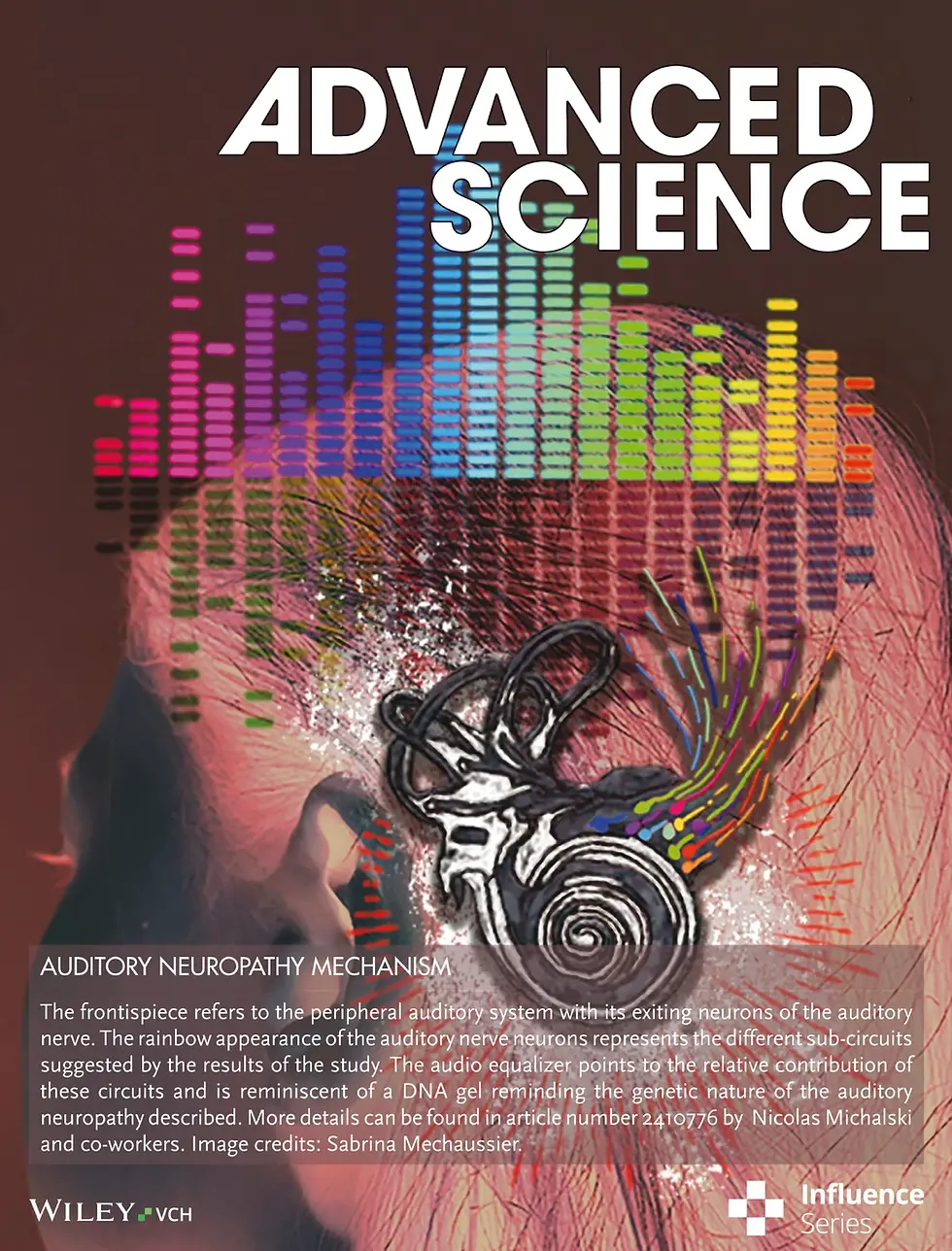
These works are highlighted on the cover of the revue.
Links :
Awake perception is associated with dedicated neuronal assemblies in cerebral cortex, Nature Neuroscience, 28 septembre 2022 Anton Filipchuk1, Joanna Schwenkgrub2, Alain Destexhe1*†, Brice Bathellier1,2,*† 1 Department for Integrative and Computational Neuroscience (ICN), Paris-Saclay Institute of Neuroscience (NeuroPSI), UMR9197 CNRS/Université Paris-Saclay, Campus CEA, 151 Rte de la Rotonde, 91400 Saclay 2 Institut Pasteur, Université de Paris, INSERM, Institut de l’Audition, 63 rue de Charenton, F-75012 Paris, France. *Corresponding author. Email: brice.bathellier at cnrs.fr, alain.destexhe at cnrs.fr † These authors contributed equally to this work
___
Activity of a neuron assembly in the auditory cortex during wakefulness (white dots) and under anesthesia (green dots). Each dot corresponds to an electrical impulse from a neuron. An image of neuron cell bodies is superimposed on the diagram. © Joanna Schwenkgrub, Institut Pasteur ; Anton Filipchuk, CNRS – Institut Pasteur


